Ct scan abdomen anatomy pdf Ballantrae

CT Anatomy of the Upper Abdomen med.wayne.edu scanner abdominal (Repères anatomiques) Dr Fourati M, Techniques d’acquisition et d’interprétation Plan de lecture d’un scanner abdominal Anatomie TDM du foie et des voies biliaires Anatomie TDM du pancréas Anatomie TDM de la rate Anatomie TDM des reins et des surrénales Anatomie TDM de l’appendice. Technique d’acquisition Scanner multicoupes+++ Rapidité d’acquisition
A Systematic Approach to the Interpretation of CT Abdomen
Scanner abdominal principe et but - Ooreka. Normal CT of the abdomen for reference. Oral and intravenous contrast given. Scan performed supine during the portal venous phase. Previous hysterectomy. Duplex collecting system on the right side, which is an anatomical variant., Normal abdomen computed tomography of a 28 year old man. Axial [5 mm thick] images were obtained with intravenous contrast in porto-venous phase Matrix size 512x512 pixels. Coronal and sagittal 2-dimensional reconstructions were also obtained. Interslice distance 5 mm. CT windows: abdomen (WL/WW 40/290 HU), bone (WL/WW 300/1500 HU). Also provided: 3D-visualization volume rendering ….
Learning the nodal stations in the abdomen Article · Literature Review (PDF Available) in The British journal of radiology 80(958):841-8 · November 2007 with 38,259 Reads How we measure 'reads' 01/07/2019 · This tutorial takes you through the important anatomy required to understand CT images of the brain. Tutorial orientation. CT images of the brain are conventionally viewed from below, as if looking up into the top of the head.
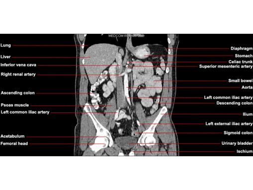
Radiology basics of abdominal CT anatomy with annotated coronal images and scrollable axial images to help medical students and junior doctors learning anatomy. Radiology basics - Abdomen anatomy … Scan for mobile link. Computed Tomography (CT) - Abdomen and Pelvis Computed tomography (CT) of the abdomen and pelvis is a diagnostic imaging test used to help detect diseases of the small bowel, colon and other internal organs and is often used to determine the cause of unexplained pain. CT scanning is fast, painless, noninvasive and accurate
Learning the nodal stations in the abdomen Article · Literature Review (PDF Available) in The British journal of radiology 80(958):841-8 · November 2007 with 38,259 Reads How we measure 'reads' Artifacts degrade the quality of images. MRI scans are more susceptible to motion artifacts than CT scans. The type of anatomy the health care provider is trying to visualize may affect their decision to order an MRI or a CT scan. For example, anatomy that contains calcium or air may not be visualized as well on an MRI scan, as compared to a CT
CT ABDOMEN ANATOMY 1. CT DR SAKHER-ALKHADERI CONSULTANT RADIOLOGIST AMC CT ABDOMEN ANATOMY 2. Cross section anatomy of abdominal ct scan 3. Anatomy of the liver segments 4. Anatomy of the liver segments 5. Right hepatic vein divides the right lobe into anterior and posterior segments. Middle hepatic vein divides the liver into right and left CT Anatomy of the Upper Abdomen - med.wayne.edu Image 1
Q1: Click on the number, that identifies liquid density. Abdomen, in human anatomy, the body cavity lying between the chest or thorax above and the pelvis below and from the spine in the back to the wall of abdominal muscles in the front. The diaphragm is its upper boundary. There is no wall or clear-cut boundary between it and the pelvis. It contains
The abdomen and pelvis contain the digestive organs as well as the urinary, endocrine, and reproductive systems. A CT scan of this area may be done to look for abscesses, tumors, kidney stones, infections, or the cause of unexplained abdominal pain. Normal abdomen computed tomography of a 28 year old man. Axial [5 mm thick] images were obtained with intravenous contrast in porto-venous phase Matrix size 512x512 pixels. Coronal and sagittal 2-dimensional reconstructions were also obtained. Interslice distance 5 mm. CT windows: abdomen (WL/WW 40/290 HU), bone (WL/WW 300/1500 HU). Also provided: 3D-visualization volume rendering …
Find the perfect ct scan abdomen stock photo. Huge collection, amazing choice, 100+ million high quality, affordable RF and RM images. No need to register, buy now! As with the systematic approach preferred for the evaluation and management of other processes explored on this site, a similarly structured method for the interpretation of imaging commonly obtained in the emergency department may afford the same benefits – namely, the timely identification of pathology while avoiding costly missed diagnoses.
24/09/2015 · Why the Test is Performed An abdominal CT scan makes detailed pictures of the structures inside your belly (abdomen) very quickly. This test may be used to look for: Cause of abdominal pain or swelling Hernia Cause of a fever Masses and tumors, including cancer Infections or … The various regions of the abdomen referred to in the description of surface anatomy and in the localisation of pathology are shown in Fig. 4.1.The lower costal margin extends from the xiphoid process of the sternum to the 10th costal cartilage (Fig. 4.2).The transpyloric plane passing across the lower border of L1 vertebra lies halfway between the suprasternal notch and the pubic symphysis.
This is a basic article for medical students and other non-radiologists. CT abdomen is an increasingly common investigation that is used to help make diagnoses of a broad range of pathologies. A CT abdomen in its simplest form is a CT from diaphragm to symphysis pubis performed 60 seconds after pump-injection of iodinated contrast into a peripheral vein. scanner abdominal (Repères anatomiques) Dr Fourati M, Techniques d’acquisition et d’interprétation Plan de lecture d’un scanner abdominal Anatomie TDM du foie et des voies biliaires Anatomie TDM du pancréas Anatomie TDM de la rate Anatomie TDM des reins et des surrénales Anatomie TDM de l’appendice. Technique d’acquisition Scanner multicoupes+++ Rapidité d’acquisition
The abdomen and pelvis contain the digestive organs as well as the urinary, endocrine, and reproductive systems. A CT scan of this area may be done to look for abscesses, tumors, kidney stones, infections, or the cause of unexplained abdominal pain. Afin que le défilement des images de scanner de l'abdomen puisse s'effectuer correctement sur l'écran ou que les légendes soient lisibles, javascript doit être activé. Flash player ou un autre plug-in n'est pas requis pour la lecture de l'atlas.
futurenergiepropres.free.fr

A Systematic Approach to the Interpretation of CT Abdomen. Find the perfect ct scan abdomen stock photo. Huge collection, amazing choice, 100+ million high quality, affordable RF and RM images. No need to register, buy now!, 01/09/2009 · The video will describe anatomical structures as seen on a CT Scan. Please see disclaimer on my website..
Local Coverage Determination for CT of the Abdomen and
Imaging Anatomy. CT und MRI Schnittbildanatomie. sectional-anatomy.org ist ein freies online Programm zur Schnittbildanatomie. mehr > CT Anatomy of the Upper Abdomen - med.wayne.edu Image 1.

Computed tomography (CT) scanning is an extremely common imaging modality in modern medicine.With advancements in technology, it is rapidly replacing many diagnostic radiographic procedures. In this article, we will outline the basic science behind CT scans, describe the principles of interpretation, and highlight their advantages and drawbacks compared to other imaging techniques. Computed tomography (CT) scanning is an extremely common imaging modality in modern medicine.With advancements in technology, it is rapidly replacing many diagnostic radiographic procedures. In this article, we will outline the basic science behind CT scans, describe the principles of interpretation, and highlight their advantages and drawbacks compared to other imaging techniques.
Abdomen and pelvis CT ANATOMY MAMDOUH MAHFOUZ MD mamdouh.m5@gmail.com www.ssregypt.com Indications Patient preparation Patient position Scanogram • To assess equivocal imaging findings • Staging of hepatic neoplasms • Metastatic workup of primary malignancies • Diagnosis of abdominal masses • Assessment of biliary problems • Diagnosis of vascular lesions • Assessment … Abdominal CT: detailled anatomy. Atlas of CT Anatomy of the Abdomen. This photo gallery presents the anatomy of the abdomen by means of CT (axial, coronal, and sagittal reconstructions).
Abdominal CT scans (also called CAT scans), are a type of specialized X-ray. They help your doctor see the organs, blood vessels, and bones in your abdomen. We’ll explain why your doctor may Artifacts degrade the quality of images. MRI scans are more susceptible to motion artifacts than CT scans. The type of anatomy the health care provider is trying to visualize may affect their decision to order an MRI or a CT scan. For example, anatomy that contains calcium or air may not be visualized as well on an MRI scan, as compared to a CT
Scan for mobile link. Computed Tomography (CT) - Abdomen and Pelvis Computed tomography (CT) of the abdomen and pelvis is a diagnostic imaging test used to help detect diseases of the small bowel, colon and other internal organs and is often used to determine the cause of unexplained pain. CT scanning is fast, painless, noninvasive and accurate Abdominal CT scan made easy 1. Abdominal CT Scan Crossing the barrier in bedside interpretation 2. DR. MASRUR AKBAR KHAN MBBS, FCPS (Surgery) 3. Can a Clinician interpret CT scan like a Radiologist? 4. OVERVIEW 5. • Introduction • CT anatomy abdomen • CT section with pathology • Take home message 6. INTRODUCTION 7.
ance ofnormal female pelvic anatomy willenable more accurate evalu-ation ofpelvic abnormalities..INTRODUCTION Thecomputed tomographic (CT)appearance ofthenormal ligamentous, vascular, and visceral anatomy ofthefemale pelvis canbeconfusing unless one isfamiliar with thebasic anatomy ofthese structures and normal variations intheir appear-ance 07/04/2019 · CT Scan Abdomen and Pelvis What is a CT scan? A CT scanner emits a series of narrow beams through the human body as it moves through an arc. …
24/09/2015 · Why the Test is Performed An abdominal CT scan makes detailed pictures of the structures inside your belly (abdomen) very quickly. This test may be used to look for: Cause of abdominal pain or swelling Hernia Cause of a fever Masses and tumors, including cancer Infections or … Normal CT of the abdomen for reference. Oral and intravenous contrast given. Scan performed supine during the portal venous phase. Previous hysterectomy. Duplex collecting system on the right side, which is an anatomical variant.
Scan for mobile link. Computed Tomography (CT) - Chest Computed tomography (CT) of the chest uses special x-ray equipment to examine abnormalities found in other imaging tests and to help diagnose the cause of unexplained cough, shortness of breath, chest pain, fever and other chest symptoms. CT scanning is fast, painless, noninvasive and accurate. Find the perfect ct scan abdomen stock photo. Huge collection, amazing choice, 100+ million high quality, affordable RF and RM images. No need to register, buy now!
Abdomen and pelvis CT ANATOMY MAMDOUH MAHFOUZ MD mamdouh.m5@gmail.com www.ssregypt.com Indications Patient preparation Patient position Scanogram • To assess equivocal imaging findings • Staging of hepatic neoplasms • Metastatic workup of primary malignancies • Diagnosis of abdominal masses • Assessment of biliary problems • Diagnosis of vascular lesions • Assessment … Computed tomography (CT) scanning is an extremely common imaging modality in modern medicine.With advancements in technology, it is rapidly replacing many diagnostic radiographic procedures. In this article, we will outline the basic science behind CT scans, describe the principles of interpretation, and highlight their advantages and drawbacks compared to other imaging techniques.
Radiology basics of abdominal CT anatomy with annotated coronal images and scrollable axial images to help medical students and junior doctors learning anatomy. Radiology basics - Abdomen anatomy … CT Anatomy of the Upper Abdomen - med.wayne.edu Image 1
Artifacts degrade the quality of images. MRI scans are more susceptible to motion artifacts than CT scans. The type of anatomy the health care provider is trying to visualize may affect their decision to order an MRI or a CT scan. For example, anatomy that contains calcium or air may not be visualized as well on an MRI scan, as compared to a CT Normal abdomen computed tomography of a 28 year old man. Axial [5 mm thick] images were obtained with intravenous contrast in porto-venous phase Matrix size 512x512 pixels. Coronal and sagittal 2-dimensional reconstructions were also obtained. Interslice distance 5 mm. CT windows: abdomen (WL/WW 40/290 HU), bone (WL/WW 300/1500 HU). Also provided: 3D-visualization volume rendering …
CT ABDOMEN ANATOMY 1. CT DR SAKHER-ALKHADERI CONSULTANT RADIOLOGIST AMC CT ABDOMEN ANATOMY 2. Cross section anatomy of abdominal ct scan 3. Anatomy of the liver segments 4. Anatomy of the liver segments 5. Right hepatic vein divides the right lobe into anterior and posterior segments. Middle hepatic vein divides the liver into right and left CT ABDOMEN ANATOMY 1. CT DR SAKHER-ALKHADERI CONSULTANT RADIOLOGIST AMC CT ABDOMEN ANATOMY 2. Cross section anatomy of abdominal ct scan 3. Anatomy of the liver segments 4. Anatomy of the liver segments 5. Right hepatic vein divides the right lobe into anterior and posterior segments. Middle hepatic vein divides the liver into right and left
CT Anatomy of the Upper Abdomen med.wayne.edu

(PDF) Learning the nodal stations in the abdomen. Anatomy of the abdominal cavity and the male pelvis: how to view anatomical labels. This tool provides access to a CT atlas in the axial plane, allowing the user to interactively learn abdominal anatomy. The images are labeled, providing an invaluable medical tool. …, 8. Is this a normal scan? This CT scan has been performed without contrast - in this case, it is a KUB scan that was performed to look for renal stones. Did you find the stone? It is in the left ureter at the level of the pelvic brim. There is associated mild hydroureter and hydronephrosis on this side..
A Systematic Approach to the Interpretation of CT Abdomen
CTAnatomy of theFemale Pelvis ASecond Look1. Radiology basics of abdominal CT anatomy with annotated coronal images and scrollable axial images to help medical students and junior doctors learning anatomy. Radiology basics - Abdomen anatomy …, Afin que le défilement des images de scanner de l'abdomen puisse s'effectuer correctement sur l'écran ou que les légendes soient lisibles, javascript doit être activé. Flash player ou un autre plug-in n'est pas requis pour la lecture de l'atlas..
Learning the nodal stations in the abdomen Article · Literature Review (PDF Available) in The British journal of radiology 80(958):841-8 · November 2007 with 38,259 Reads How we measure 'reads' 01/07/2019 · This tutorial takes you through the important anatomy required to understand CT images of the brain. Tutorial orientation. CT images of the brain are conventionally viewed from below, as if looking up into the top of the head.
Anatomy of the abdominal cavity and the male pelvis: how to view anatomical labels. This tool provides access to a CT atlas in the axial plane, allowing the user to interactively learn abdominal anatomy. The images are labeled, providing an invaluable medical tool. … Computed Tomography (CT) - Abdomen and Pelvis What is CT Scanning of the Abdomen/Pelvis? CT scanning—sometimes called CAT scanning—is a noninvasive medical test that helps physicians diagnose and treat medical conditions. CT scanning combines special x …
Abdominal CT: detailled anatomy. Atlas of CT Anatomy of the Abdomen. This photo gallery presents the anatomy of the abdomen by means of CT (axial, coronal, and sagittal reconstructions). CT ABDOMEN ANATOMY 1. CT DR SAKHER-ALKHADERI CONSULTANT RADIOLOGIST AMC CT ABDOMEN ANATOMY 2. Cross section anatomy of abdominal ct scan 3. Anatomy of the liver segments 4. Anatomy of the liver segments 5. Right hepatic vein divides the right lobe into anterior and posterior segments. Middle hepatic vein divides the liver into right and left
ance ofnormal female pelvic anatomy willenable more accurate evalu-ation ofpelvic abnormalities..INTRODUCTION Thecomputed tomographic (CT)appearance ofthenormal ligamentous, vascular, and visceral anatomy ofthefemale pelvis canbeconfusing unless one isfamiliar with thebasic anatomy ofthese structures and normal variations intheir appear-ance Abdominal CT Atlas. Talos I-F., Jakab M., Kikinis R. SPL Abdominal Atlas. SPL 2015 Sep;
Computed tomography of the abdomen and pelvis is an application of computed tomography (CT) and is a sensitive method for diagnosis of abdominal diseases. It is used frequently to determine stage of cancer and to follow progress. It is also a useful test to investigate acute abdominal pain (especially of the lower quadrants, whereas ultrasound is the preferred first line investigation for ONLINE MRI & CT SECTIONAL ANATOMY Kenneth K. F. Ho Bachelor of Medicine, Bachelor of Surgery (University of Hong Kong) Fellow, Hong Kong College of Radiologists Fellow, Hong Kong Academy of Medicine (Radiology) Real-time interface human sectional anatomy. Welcome to Online MRI & CT Sectional Anatomy. Online MRI & CT Sectional Anatomy (OMCSA K-anatomy) is probably one of the …
Afin que le défilement des images de scanner de l'abdomen puisse s'effectuer correctement sur l'écran ou que les légendes soient lisibles, javascript doit être activé. Flash player ou un autre plug-in n'est pas requis pour la lecture de l'atlas. CT abdomen general Indication/Technique. Please see the X-ray/CT Technique class for additional information about the technique of ‘computer tomography’ (CT). Abdominal CT scans are generally evaluated in the transversal direction; the patient is seen from the feet upward as it were.
CT abdomen general Indication/Technique. Please see the X-ray/CT Technique class for additional information about the technique of ‘computer tomography’ (CT). Abdominal CT scans are generally evaluated in the transversal direction; the patient is seen from the feet upward as it were. 01/07/2019 · This tutorial takes you through the important anatomy required to understand CT images of the brain. Tutorial orientation. CT images of the brain are conventionally viewed from below, as if looking up into the top of the head.
The abdomen and pelvis contain the digestive organs as well as the urinary, endocrine, and reproductive systems. A CT scan of this area may be done to look for abscesses, tumors, kidney stones, infections, or the cause of unexplained abdominal pain. Abdominal CT The CT of the abdomen extends from the dome of the diaphragm to the pelvic brim or pubic symphysis, depending upon whether one groups the pelvis with the abdomen or treats it separately. A CT scan of the abdomen will be considered medically reasonable and necessary under the following Printed on 11/5/2018. Page 2 of 113
Computed Tomography (CT) - Abdomen and Pelvis What is CT Scanning of the Abdomen/Pelvis? CT scanning—sometimes called CAT scanning—is a noninvasive medical test that helps physicians diagnose and treat medical conditions. CT scanning combines special x … Computed tomography (CT) scanning is an extremely common imaging modality in modern medicine.With advancements in technology, it is rapidly replacing many diagnostic radiographic procedures. In this article, we will outline the basic science behind CT scans, describe the principles of interpretation, and highlight their advantages and drawbacks compared to other imaging techniques.
Qu’est-ce qu’un scanner abdomino-pelvien ? Cet examen permet de faire des images en coupe de l’ensemble des organes de l’abdomen. Le scanner utilise des rayons X. En matière d’irradiation des patients, rien n’a pu être démontré dans ce domaine compte tenu des faibles doses utilisées et des précautions prises pour limiter au strict minimum la zone examinée. Computed tomography (CT) scanning is an extremely common imaging modality in modern medicine.With advancements in technology, it is rapidly replacing many diagnostic radiographic procedures. In this article, we will outline the basic science behind CT scans, describe the principles of interpretation, and highlight their advantages and drawbacks compared to other imaging techniques.
Anatomy of the thorax (CT)
CT Anatomy of the Upper Abdomen med.wayne.edu. Qu’est-ce qu’un scanner abdomino-pelvien ? Cet examen permet de faire des images en coupe de l’ensemble des organes de l’abdomen. Le scanner utilise des rayons X. En matière d’irradiation des patients, rien n’a pu être démontré dans ce domaine compte tenu des faibles doses utilisées et des précautions prises pour limiter au strict minimum la zone examinée., Learning the nodal stations in the abdomen Article · Literature Review (PDF Available) in The British journal of radiology 80(958):841-8 · November 2007 with 38,259 Reads How we measure 'reads'.
Computed Tomography (CT) Chest - RadiologyInfo.org. Abdominal CT: detailled anatomy. Atlas of CT Anatomy of the Abdomen. This photo gallery presents the anatomy of the abdomen by means of CT (axial, coronal, and sagittal reconstructions)., This is a basic article for medical students and other non-radiologists. CT abdomen is an increasingly common investigation that is used to help make diagnoses of a broad range of pathologies. A CT abdomen in its simplest form is a CT from diaphragm to symphysis pubis performed 60 seconds after pump-injection of iodinated contrast into a peripheral vein..
CT ABDOMEN ANATOMY SlideShare

Scanner abdomino-pelvien C.S.E. Imagerie. The abdomen and pelvis contain the digestive organs as well as the urinary, endocrine, and reproductive systems. A CT scan of this area may be done to look for abscesses, tumors, kidney stones, infections, or the cause of unexplained abdominal pain. 07/04/2019 · CT Scan Abdomen and Pelvis What is a CT scan? A CT scanner emits a series of narrow beams through the human body as it moves through an arc. ….
Abdomen and pelvis CT ANATOMY MAMDOUH MAHFOUZ MD mamdouh.m5@gmail.com www.ssregypt.com Indications Patient preparation Patient position Scanogram • To assess equivocal imaging findings • Staging of hepatic neoplasms • Metastatic workup of primary malignancies • Diagnosis of abdominal masses • Assessment of biliary problems • Diagnosis of vascular lesions • Assessment … Abdominal CT scans (also called CAT scans), are a type of specialized X-ray. They help your doctor see the organs, blood vessels, and bones in your abdomen. We’ll explain why your doctor may
CT abdomen general Indication/Technique. Please see the X-ray/CT Technique class for additional information about the technique of ‘computer tomography’ (CT). Abdominal CT scans are generally evaluated in the transversal direction; the patient is seen from the feet upward as it were. The abdomen and pelvis contain the digestive organs as well as the urinary, endocrine, and reproductive systems. A CT scan of this area may be done to look for abscesses, tumors, kidney stones, infections, or the cause of unexplained abdominal pain.
Abdominal CT Atlas. Talos I-F., Jakab M., Kikinis R. SPL Abdominal Atlas. SPL 2015 Sep; Abdominal CT scans (also called CAT scans), are a type of specialized X-ray. They help your doctor see the organs, blood vessels, and bones in your abdomen. We’ll explain why your doctor may
Chest, Abdomen and Pelvis CT Protocols Chest Chest CT Low Dose Nodule Evaluation Chest CT Lung Cancer Screening Chest CT Routine With Contrast Chest CT Without Routine Chest CTA- Pulmonary Embolism Chest CTA- Acute Aorta Chest CTA- Aortic Aneurysm -Pre EVT Chest CTA Aortic Aneurysm- Post EVT Chest CTA- Acute Aorta- Trauma Chest CT High Resolution Coronary CTA Screening History Chest, Abdomen Abdominal CT: detailled anatomy. Atlas of CT Anatomy of the Abdomen. This photo gallery presents the anatomy of the abdomen by means of CT (axial, coronal, and sagittal reconstructions).
CT abdomen general Indication/Technique. Please see the X-ray/CT Technique class for additional information about the technique of ‘computer tomography’ (CT). Abdominal CT scans are generally evaluated in the transversal direction; the patient is seen from the feet upward as it were. Abdominal CT: detailled anatomy. Atlas of CT Anatomy of the Abdomen. This photo gallery presents the anatomy of the abdomen by means of CT (axial, coronal, and sagittal reconstructions).
01/07/2019 · This tutorial takes you through the important anatomy required to understand CT images of the brain. Tutorial orientation. CT images of the brain are conventionally viewed from below, as if looking up into the top of the head. Normal CT of the abdomen for reference. Oral and intravenous contrast given. Scan performed supine during the portal venous phase. Previous hysterectomy. Duplex collecting system on the right side, which is an anatomical variant.
Q1: Click on the number, that identifies liquid density. Abdomen and pelvis CT ANATOMY MAMDOUH MAHFOUZ MD mamdouh.m5@gmail.com www.ssregypt.com Indications Patient preparation Patient position Scanogram • To assess equivocal imaging findings • Staging of hepatic neoplasms • Metastatic workup of primary malignancies • Diagnosis of abdominal masses • Assessment of biliary problems • Diagnosis of vascular lesions • Assessment …
ance ofnormal female pelvic anatomy willenable more accurate evalu-ation ofpelvic abnormalities..INTRODUCTION Thecomputed tomographic (CT)appearance ofthenormal ligamentous, vascular, and visceral anatomy ofthefemale pelvis canbeconfusing unless one isfamiliar with thebasic anatomy ofthese structures and normal variations intheir appear-ance Abdominal CT scan made easy 1. Abdominal CT Scan Crossing the barrier in bedside interpretation 2. DR. MASRUR AKBAR KHAN MBBS, FCPS (Surgery) 3. Can a Clinician interpret CT scan like a Radiologist? 4. OVERVIEW 5. • Introduction • CT anatomy abdomen • CT section with pathology • Take home message 6. INTRODUCTION 7.
Scan for mobile link. Computed Tomography (CT) - Abdomen and Pelvis Computed tomography (CT) of the abdomen and pelvis is a diagnostic imaging test used to help detect diseases of the small bowel, colon and other internal organs and is often used to determine the cause of unexplained pain. CT scanning is fast, painless, noninvasive and accurate ONLINE MRI & CT SECTIONAL ANATOMY Kenneth K. F. Ho Bachelor of Medicine, Bachelor of Surgery (University of Hong Kong) Fellow, Hong Kong College of Radiologists Fellow, Hong Kong Academy of Medicine (Radiology) Real-time interface human sectional anatomy. Welcome to Online MRI & CT Sectional Anatomy. Online MRI & CT Sectional Anatomy (OMCSA K-anatomy) is probably one of the …
CT und MRI Schnittbildanatomie. sectional-anatomy.org ist ein freies online Programm zur Schnittbildanatomie. mehr > ance ofnormal female pelvic anatomy willenable more accurate evalu-ation ofpelvic abnormalities..INTRODUCTION Thecomputed tomographic (CT)appearance ofthenormal ligamentous, vascular, and visceral anatomy ofthefemale pelvis canbeconfusing unless one isfamiliar with thebasic anatomy ofthese structures and normal variations intheir appear-ance
Anatomy of the chest: how to view the anatomical labels. This atlas is a comprehensive and affordable learning tool for medical students and residents and especially for radiologists and pneumologists. It provides access to CT images in the axial plane, allowing the user to learn and review the lung anatomy interactively. Anatomical structures The abdomen and pelvis contain the digestive organs as well as the urinary, endocrine, and reproductive systems. A CT scan of this area may be done to look for abscesses, tumors, kidney stones, infections, or the cause of unexplained abdominal pain.